Running with Knee Osteoarthritis-Part 1

By Lillian Wynn PT, DPT
Physical Therapist

Background

40% of American adults (110 million people) report walking or running as part of a regular exercise routine.  Reports and ‘common knowledge’ about running and its impact on our joints are often conflicting. This is the first of three blog posts designed to look at current medical research regarding running on aging joints.

Article summary

Often of most concern with running is whether the impact is harmful to the knee joint, as the thought is impact could cause and/or worsen osteoarthritis. Osteoarthritis is the term given to changes that occur along a joints surface as we age. The most common way to diagnose osteoarthritis is with an x-ray. A prospective study published in The American Journal for Preventative Medicine investigated whether running as we age increases the severity or frequency of knee arthritis.

PARTICIPANTS

45 long distance runners who were 50 years old or older, and had been running for at least 10 years; and 53 controls who were 50 years or older and did not run for exercise.

METHODS

Initial x-rays were taken of both knees of all participants. Over the next 18 years, 5 follow up x-rays were taken of each patient. These x-rays were graded on a standard scale to quantify the severity of knee arthritis.

RESULTS

Runners did not show higher rates or more severe cases of knee osteoarthritis than non-runners

CONCLUSIONS

Models found that higher BMI, higher initial damage on x-ray, and age to be most strongly correlated with arthritis on x-ray. There was no data to suggest that running, gender, previous knee injury, or total exercise time contributed to osteoarthritis of the knee. In short-go out and go for your run!

 PTF approach

Here at PTF, we want to keep you active in the activities that matter to you. If walking and running are important to you, and you feel limited by your knees, an evaluation could be useful. Often tight and/or weak muscles, stiff joints, and poor movement patterns can contribute to pain while running. PTF does a complete evaluation and then designs a treatment plan individual to you and your body to keep you moving.

Original Article

Chakravarty, E., Hubert, H., Lingala, V., Zatarain, E., Fries, J. (2008). Long Distance Running and Knee Osteoarthritis A Prospective Study. American Journal of Preventative Medicine, 35(2), 133-138. doi:10.1016/j.amepre.2008.03.032.

Pilates Training May Be Your Answer to Improve Your 5K Run Performance

by Genevieve Bland, PT, DPT

Background

Running performance depends on several variables, which include: high maximum oxygen consumption, the ability to sustain maximum oxygen consumption for long periods of time, and the ability to move efficiently. The ability to move efficiently depends on metabolic cost, which is the amount of oxygen spent to move a runner a certain distance at a submaximal intensity. The lower the metabolic cost results in less energy expenditure and lower oxygen consumption, which makes a more efficient runner. There are numerous ways to lower metabolic cost such as: endurance training, strength and aerobic training, and plyometric training. Another way to improve metabolic cost is by engaging the muscles of the trunk and lower limbs to establish better control during a run. Pilates training has been utilized to strengthen trunk muscles in correlation with breathing. Research suggests, Pilates training strengthen core musculature, which decreases the amount of activation during running and increases the runner’s efficiency thereby improving running performance.

Literature search and data analysis to improve a recreational runners speed

In a recent search Finatto et al. investigated the effects of strength training of the postural and trunk muscles. There is research indicating that strength training improves running efficiency and performance that incorporate maximal and explosive programs to the lower extremities.
Finatto et al. hypothesized that metabolic cost and trunk muscle activation will be reduced and consequently, running performance may be improved. They studied the effects of strengthening the muscles of the center of force by Pilates training on metabolic cost and the muscle activation pattern and biomechanical parameters that could improve metabolic cost, which would lead to improve running performance.

Methods

Two groups
1. Pilates group: underwent running training combined with Pilates training
2. Control group: running training only

Both groups trained for 12 weeks and were evaluated before and after the training period. Post-training evaluations were performed 72 hours after the last training session

Running training: both groups participated in a 12-week racetrack training program 2x/week.

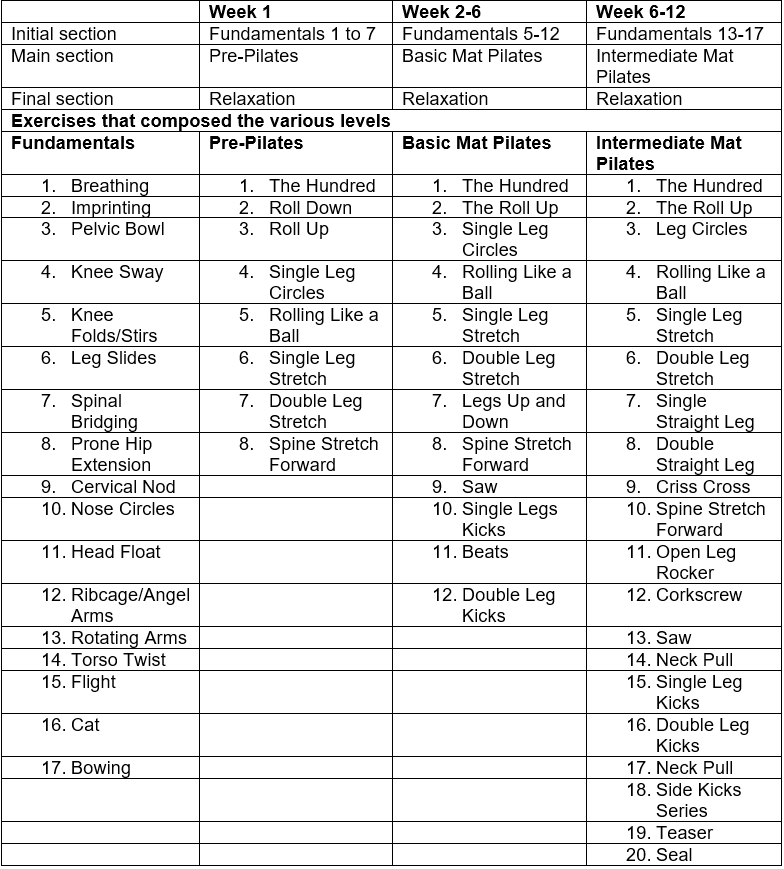
Pilate’s training: classic mat Pilates training two one-hour weekly sessions performed on days alternate to the days of the running training for the Pilates group only.

Table 1. 12-week periodization of Pilates training

12 week periodization of Pilates training

During the second session, the participants were evaluated for the maximum isometric amplitude of the electromyographic (EMG) signal of trunk muscles which include: obliquus externus, obliquus internus, longissimus, bicep femoris, vastus lateralis, latissimus dorsi, and gluteus medius. Each participant exerted max force with isometric testing after walking for 5 minutes. Three measurements were taken pre and post training. EMG were performed during running as well. Metabolic cost was measured after treadmill testing and participants rested for 15 minutes in the sitting position and at rest for five minutes in the orthostatic position to determine resting heart rate and maximal oxygen consumption.

Results

Running performance and respiratory variables were not significantly different between the groups in the pre-training period. In the post-training period, the Pilates group had significantly higher maximal oxygen consumption and significantly shorter 5-km running time with a significant lower metabolic cost. This suggests that distance runners are able to transfer effective gains from a slow-type core strength training method to the running movement.

Table 2

Effect of running training and running training combined with Pilates on performance and respiratory variables.

Data Represent the Mean Values (Standard Error) for 5-km Running Time, Maximum Oxygen Consumption (VO2max), Metabolic Cost at 10 km.h-1 (Cmet10), Metabolic Cost at 12 km.h-1 (Cmet12), Speed at the Second Ventilatory Threshold (VT2), and Oxygen Consumption at the Second Ventilatory Threshold (VO2 VT2).

Effect of running training and running training combined with Pilates on performance and respiratory variables

Physical Therapy First:

Here at Physical Therapy First we provide one on one hands on care for our patients to treat injuries, rehab after surgery and to help athletes optimize fitness performance. Our therapists can analyze your running technique and develop an individual treatment plan to assist you in achieving your goals for running.

https://journals.plos.org/plosone/article?id=10.1371/journal.pone.0194057

What effect does hip strengthening have on running and single-leg squatting mechanics?

by Leah Flamm, PT, DPT

Looking at a study by Richard W. Willy, PT, PhD, OCS, and Irene S. Davis, PT, PhD, FAPTA

Background

Abnormal hip and knee mechanics may be related to a number of running-related injuries, from stress fractures, to IT band syndrome, to patellofemoral pain syndrome (PFPS). PFPS has been linked to abnormal knee movements like excessive hip adduction, hip internal rotation, contralateral pelvic drop (when the opposite side of the pelvis drops), and knee external rotation during running, single-leg squats, single-leg jump landing, and step-downs. Studies suggest that women with PFPS may have weak hip muscles, and though hip strengthening programs have been shown to improve strength, it is not clear whether hip strengthening programs improve abnormal hip and knee mechanics while running and squatting. Though studies have looked at hip strengthening in healthy active females with normal mechanics, only two have looked at that of individuals with abnormal mechanics.

This study by Drs. Willy and Davis aimed to examine the effect of a hip-strengthening program that included movement training for the single-leg squat on hip and knee mechanics during running and squatting in females who exhibited abnormal mechanics during running.

Hypothesis

The researchers hypothesized that peak hip adduction, hip internal rotation, contralateral pelvic drop, and knee external rotation would be reduced during single-leg squats due to the specific neuromuscular reeducation of that activity.

Methods

The researchers included female runners ages 18 – 35, who were running at least 10 km per week, and were required to not do any lower extremity resistance training for at least 90 days before the study. They were assigned to either the control group or the treatment group and were not told which group they had been assigned to. The researchers measured peak hip adduction (HADD) during running and squatting because excessive hip adduction is related to lower extremity injuries in runners. The researchers measured baseline hip abduction and external rotation strength at baseline and after a strengthening program or after the control group.

Physical therapists supervised the exercise progression and ensured participants were activating the hip abductors and hip external rotators. Exercises included side-lying hip extension and external rotation, squats with resistance band targeting hip external rotators, hip hikes against the wall, side-stepping with a resistance band to target hip abductors, and then single leg squats with resistance bands targeting hip abductors.

Results

Results show that the treatment group showed significantly improved hip abduction and external rotation strength, and the control group did not. Though the running data did not show any difference in peak hip adduction between groups or between pre- and post-training, single-leg squat data did show significant improvement in peak hip adduction, hip internal rotation, and contralateral (opposite side) pelvic drop.

Discussion and conclusion

The researchers found that their hip abductor/external rotator strength training program was effective at changing hip mechanics during a single-leg squat. However, no changes in running mechanics were noted, which suggests that strengthening the hip muscles alone may not be enough to change movement patterns while running.

Why go to a physical therapist?

A physical therapist (PT) can examine and evaluate an individual’s mechanics while running, squatting, or performing any physical activity. Then the PT can determine what weaknesses and movement patterns may be present. Physical therapists can provide exercises to strengthen and stretch weak and tight muscles, but they can also work on retraining movement patterns that may be associated with injuries. Pure strengthening may not be enough to change the mechanics that may be underlying some injuries in athletes or anyone. For that reason, we may also focus on neuromuscular reeducation to change movement patterns so that we can address the root cause of the problem.

Original article

Willy, R. W., & Davis, I. S. (2011). The Effect of a Hip-Strengthening Program on Mechanics During Running and During a Single-Leg Squat. Journal of Orthopaedic & Sports Physical Therapy,41(9), 625-632. doi:10.2519/jospt.2011.3470

ACL Injury

by Scott Vocke, DPT, CMTPT
Physical Therapist

The ACL is one of four ligaments that helps to stabilize the knee. It is most commonly injured during quick stopping and twisting motions of the knee with the foot planted on the ground. Surgery can be performed to repair or reconstruct the damaged ligament. In many cases, ACL surgery is the best option for treatment. This is especially true for athletes that participate in physically demanding sports that involve cutting and pivoting movements, which require an intact ACL for optimal knee stability. However, research suggests in many instances nonoperative management may be a better option.

Research for ACL Operative vs. Non-operative Management

In a systematic review published in “The Knee” journal (an Official Publication of the British Association for Surgery of the Knee), researchers presented evidence that suggests ACL reconstruction surgery for a specific group of patients may not be any more beneficial than conservative management (also known as rehabilitation without surgery).1 Research has also been conducted to identify two categories of patients with ACL injury: “copers” and “non-copers.”2 Patients who are considered copers are individuals who are able to perform functional activities despite an ACL rupture. Non-copers are patients who are unable to perform functional activities and have repetitive episodes of the knee “giving-way” (also known as instability). These non-copers are less likely candidates for non-operative treatment. There are many evidence-based assessment tools that physical therapists can use to help determine if a patient is a coper or non-coper, thereby indicating whether conservative management is a potential treatment for their injury.

Research for Exercise to Improve Knee Function Early After ACL Injury(3)

A study published in the Journal of Orthopedic Sports Physical Therapy investigated a progressive five-week exercise therapy program early after ACL injury with the following goals:(3)
• Present a progressive exercise program that can be used for patients with ACL injury.
• Evaluate changes in quadricep and hamstring muscle strength
• Assess dynamic lower extremity muscular power and strength compared to the unaffected leg with validated single leg hop tests
• Determine changes in knee function based on self-assessment tools
• Study the difference between coper and non-copers after completion of the exercise program
• Examine potential risk of adverse events from an intensive exercise program early after ACL injury

Researchers hypothesized that patients would:

• Improve knee function based on strength measurements, hop test outcomes, and knee function self-assessment scores
• Non-copers would improve knee function more than copers
• Patients would not have any adverse events during the progressive therapy exercise program.

Participants

• 100 patients 13-60 years of age
• Participate regularly in pivoting sports
• Complete ACL tear verified by MRI within the 90 days of initial visit and without evidence of other structural damage to the knee.
• Ability to participate in exercise program two times per week

Exercise program

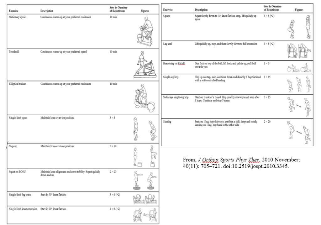
The exercise program was started as soon as knee joint swelling was eliminated and full range of motion was restored. Subjects participated in the exercise program two to four times per week, which included intensive muscle strength training, plyometric exercises and neuromuscular re-education exercises. Dosage of exercise was based on recommendations form the American College of Sports Medicine and the specific exercises from the program are identified in the following diagram from the research article:

ACL Injury Exercise

Outcomes

Findings from the study showed significant increases in knee function, increased hamstring and quad strength, and improved single leg hop test performance in both coper and non-coper groups. Based on these results, researchers concluded that the progressive exercise therapy program was low risk for adverse events. Therefore, the program should be used for non-operative management of ACL tears as well as to improve knee function prior to ACL surgery.

Physical Therapy First Approach

Determining if a patient is a candidate for non-operative management of an ACL injury is a multidisciplinary approach and may include an orthopedic surgeon. If non-operative management of an ACL injury is determined to be the best option for a patient with an ACL injury, he/she will undergo a full physical therapy evaluation to determine impairments and functional limitations.

These impairments and limitations will be addressed with evidence-based treatments, such as the exercise program from the article reviewed above. Physical therapy treatments to help recover from an ACL injury may include:

• Joint mobilization
• Soft tissue mobilization
• Specific muscle strengthening
• Neuromuscular re-education interventions
• Balance training
• Muscle stretching
• Correction of biomechanical faults in functional movement
• Home exercise program development
• Physical therapy modalities, such as, moist heat, cold packs, ultrasound, electrical stimulation, phototherapy/laser

References

1. Smith TO, Postle K, Penny F, McNamara I, Mann CJ. Is reconstruction the best management strategy for anterior cruciate ligament rupture? A systematic review and meta-analysis comparing anterior cruciate ligament reconstruction versus non-operative treatment. Knee. 2014;21:462–70. [PubMed]
2. Fitzgerald GK, Axe MJ, Snyder-Mackler L. A decision-making scheme for returning patients to high-level activity with nonoperative treatment after anterior cruciate ligament rupture. Knee Surg Sports Traumatol Arthrosc. 2000;8:76–82. doi: 10.1007/s001670050190. [PubMed]
3. Eitzen I, Moksnes H, Snyder-Mackler L, Risberg MA. A Progressive 5-Week Exercise Therapy Program Leads to Significant Improvement in Knee Function Early After Anterior Cruciate Ligament Injury. Orthop Sports Phys Ther. 2010 November ; 40(11): 705–721. doi:10.2519/jospt.2010.3345.

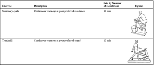
Stationary Cycle and Treadmill

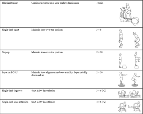
ACL Excercises

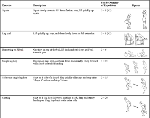
ACL Exercises

What are the Risk Factors for Reinjury Following an ACL Reconstruction?

by Alex Tan, PT, DPT OCS
Physical Therapist

Background:

Anterior cruciate ligament (ACL) rupture rate is the highest in young athletes who participate in sports involving cutting and pivoting (basketball, skiing, soccer) and when they occur tend to be season-ending injuries requiring surgical reconstruction. Female sex and young age have been viewed as common risk factors for initial injury. Even after successful surgical reconstruction and rehabilitation, reinjury to the reconstructed ACL and/or ACL of the opposite limb can occur. At this time, research suggests that environmental and genetic factors may be causes of reinjury. Recently, more studies have become available focusing on the rates of reinjury in younger active individuals, but their results have not been summarized.

Literature search and data analysis to determine possible risk factors for reinjury:

In a recent article by Wiggins et al researchers reviewed existing literature and analyzed data found within to evaluate whether a patient’s age and activity level could be seen as risk factors for another ACL injury following reconstruction.

The researchers narrowed down a database search to 19 articles for review. Then they recorded data from populations included in these articles regarding: total ACL reinjury rate (of the same side and/or opposite limb), specific sport an individual returned to if provided, the nature of the sport (low risk or high-risk involving cutting/jumping), and demographics.

The researchers hypothesized that returning to sport and a younger age would place individuals at higher risk for a second ACL injury.

An analysis was performed on the data from the entire included population and a separate analysis was performed on data sets broken down by individuals of a young age, those who returned to a sport, and from those who were young and returned to a sport.

Results

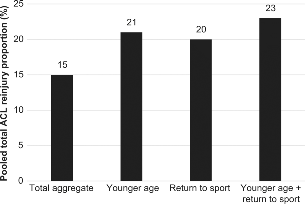
  • In individuals across all ages who underwent ACL reconstruction: 7% reinjured the same ACL and 8% reinjured the opposite side
  • In individuals < 25 years old who underwent ACL reconstruction: 21% had another ACL injury with 11% being the opposite knee
  • In individuals who returned to sports following reconstruction: 20% had another ACL injury with 12% being the opposite knee
  • In individuals who were <25 years old who returned to high-risk sport following reconstruction: 23% had another injury with 12%

ACL Reinjury Data Graph

Conclusion

Individuals who return to a high-risk sport and those of a younger age are at risk for ACL reinjury. Almost one-fourth of individuals who are of a younger age (<20-25) and return to a high risk sport that involves pivoting and cutting will either reinjure the reconstructed ACL or injure the opposite side. Based on the current literature the majority of these secondary injuries occur in the knee that has not undergone previous surgical intervention.

What Can Physical Therapists Do to Prevent Reinjury?

Individuals who have or have not undergone a previous ACL repair can be examined by a physical therapist who can determine if they are at risk for injury/reinjury due to factors such as:

  • The individual’s improper performance of functional movements
  • Muscular imbalances
  • Strength and flexibility deficits
  • Improper running and landing mechanics
  • Balance deficits
  • Asymmetrical strength/power between limbs

Your therapist will then address these issues with a personalized plan of care which may include but is not limited to interventions such as:

  • Exercises to strengthen or improve flexibility in specific muscles
  • Plyometric or jump training
  • Activity modification recommendations
  • Neuromuscular re-education
  • Bracing

Original Article: Wiggins Amelia, Grandhi Ravi, Schneider Daniel, Stanfield Denver, Webster Kate, Myer Gregory. Risk of secondary injury in younger athletes after anterior cruciate ligament reconstruction: A systematic review and meta-analysis Am J. Sports Med. 2016 July; 44(&): 1861-1876

https://www.ncbi.nlm.nih.gov/pmc/articles/PMC5501245/

Physical Therapy and Online Programs to treat Hip Osteoarthritis, Knee Osteoarthritis, or Both

by John A. Baur, PT, DPT, OCS, CSCS, FAAOMPT
Physical Therapist

Many people are finding information about treatments for health conditions on the internet. A novel approach to physical therapy takes advantage of this propensity to engage online with treatment. This blended approach combines face-to-face visits with an online program or e-Exercise. But is this as effective as traditional physical therapy?

Comparing a Blended Approach with Traditional Physical Therapy

Researchers in a recent study[i] compared the short and long-term effectiveness of a blended approach, which uses an online e-Exercise program to the traditional approach of physical therapy alone. They focused on patients with osteoarthritis of the knee and/or hip. This condition is the most common chronic condition of the joints, causing pain, stiffness and swelling.

Research Participants

The study enrolled 208 patients selected from 143 physical therapy practices. Patients had to meet the following criteria:

· 40 – 80 years of age

· Osteoarthritis of the hip and/or knee

· Not on a waiting list for hip or knee replacement surgery

· No contra-indications for physical activity without supervision

· Moderately physically active

· No current physical therapy program in the last 6 months

· Access to the internet

Blended Approach

This approach combines physical therapy with an online application. It involves 5 face-to-face sessions with a physical therapist and enrollment in an e-Exercise program that has a graded activity module, exercises and information modules. Once the patient is enrolled in the online, e-Exercise program, the physical therapist can adapt the program to the patient’s needs and monitor login frequencies and assignment evaluation. They can then discuss progress during the face-to-face physical therapy session.

Traditional Approach

The traditional approach to treating osteoarthritis over a period of 12 months involves 12 face-to-face sessions with a physical therapist. During those sessions the physical therapist will share information about the disease and treatment and work the patient on a program of physical exercise and strength and stability exercises.

Results

Patients were assessed at 3 and 12 months on quality of life, physical functioning and physical activity. The assessment used an online questionnaire.

At 12 months there was no difference between the groups on health-related quality of life, physical functioning or physical activity. Both approaches achieved the same results.

Recommendations

When choosing between a blended approach that incorporates e-Exercise and traditional physical therapy, the preferences and predisposition of the patient should be considered. The blended approach requires more motivation on the part of the patient and they must take a more active role for the treatment to be successful. If the blended approach is used, it may also be wise to incorporate a personal activity tracker to keep patients motivated and on task.

Physical Therapy First Approach – Treatment for Osteoarthritis

At Physical Therapy First each patient undergoes a complete examination to determine the underlying biomechanical cause of her/his knee/hip osteoarthritis. Physical therapy can help knee/hip osteoarthritis and it all starts with a thorough examine which will include assessing:

– muscle imbalances

– specific muscle weakness

– muscle flexibility

– joint mobility

– shoe wear assessment

– functional movement

– posture

– neurological exam

Based on the finding from the initial physical therapy assessment, a custom treatment plan will be designed to restore movement quality and efficiently, and ultimately decreasing the pain resulting from knee/hip osteoarthritis.

Some of the treatments that may be provided in physical therapy to help address patients with osteoarthritis includes:

– individualized online/e-Exercise program

– adaptive equipment

– bracing

– proper shoe wear

– joint mobilization

– soft tissue mobilization

– specific muscle strengthening

– muscle stretching

– postural correction

– correction of biomechanical faults in functional movement

– physical therapy modalities, such as, moist heat, cold packs, ultrasound, electrical stimulation, phototherapy/laser

[i] Corelien J J Kloek, Daniël Bossen, Peter M Spreeuwenberg, Joost Dekker, Dinny H de Bakker, Cindy Veenhof; Effectiveness of a Blended Physical Therapist Intervention in People With Hip Osteoarthritis, Knee Osteoarthritis, or Both: A Cluster-Randomized Controlled Trial, Physical Therapy, Volume 98, Issue 7, 1 July 2018, Pages 560–570, https://doi.org/10.1093/ptj/pzy045