by ptfadmin | Mar 9, 2026 | Health Tips
Reviewed by Tyler Tice, PT, DPT, OCS, ATC
This systematic review evaluated the clinical and preclinical evidence for cannabidiol (CBD), excluding THC, as a treatment for pain. The study followed PRISMA guidelines and searched PubMed and Web of Science using the terms “((cannabidiol) NOT (THC)) NOT (tetrahydrocannabinol)) AND (pain treatment)”. Inclusion criteria consisted of clinical and preclinical trials (in vitro and in vivo) in which CBD was used alone or as the major component of cannabis in any pharmaceutical form and free of THC. A total of 40 studies met inclusion criteria, including 11 human clinical trials, 2 canine clinical trials, and 27 preclinical studies. Exclusion criteria included the presence of THC or other cannabis components, systematic or narrative reviews, meta-analyses, case reports, and articles not published in English or Spanish. Literature screening was conducted independently by two researchers and supervised by a third reviewer
Cannabis (hemp) has been used for thousands of years for textile, medicinal, and culinary purposes. The primary compounds of pharmaceutical interest are cannabinoids, with CBD demonstrating a wide range of pharmacologic effects, including anti-inflammatory, antioxidant, anticonvulsant, and antipsychotic properties, mediated through interactions with multiple endogenous receptors. CBD can be administered orally, topically, intravenously, or intraperitoneally. Oral administration was the most common route in clinical trials, while intraperitoneal administration predominated in preclinical studies. One canine clinical trial reported that oral CBD bioavailability was approximately 19% lower compared to other formulations, highlighting the importance of administration route when determining appropriate dosing. The authors emphasized the need for further studies to establish optimal delivery methods based on specific pain pathologies.
Among the human clinical trials, 7 of 11 studies demonstrated pain-reducing effects of CBD. Conditions showing benefit included osteoarthritis, chronic pain, neuropathic pain, bruxism, arthritis, and atopic dermatitis. Importantly, no clinical or preclinical trials reported significant adverse effects or treatment discontinuation. The review concluded that CBD is a potentially effective and safe treatment for pain, supported by its analgesic and anti-inflammatory properties, though larger, well-designed clinical trials are needed to strengthen the evidence base.
Reference:
Cásedas G, Yarza-Sancho M, López V. Cannabidiol (CBD): A Systematic Review of Clinical and Preclinical Evidence in the Treatment of Pain. Pharmaceuticals (Basel). 2024;17(11):1438. Published 2024 Oct 28. doi:10.3390/ph17111438
by ptfadmin | Mar 2, 2026 | Health Tips
Reviewed by Tyler Tice, PT, DPT, OCS, ATC
Background:
- Unilateral vestibular hypofunction( UVH) : partial or complete loss of function in one of the peripheral vestibular organa, and/or nerves
- Acute UVH: most commonly due to vestibular neuritis, but also may be due to trauma, surgical transection, ototoxic meds, or Meniere’s disease.
- Bilateral vestibular hypofunction (BVH): idiopathic in 20-51% of cases
- Common presentation/symptoms:
- dizziness, imbalance, and/or oscillopsia, gaze and gait instability, and impaired navigation and spatial orientation
- Spatial navigation impairments
- Memory, executive function, attention
- Prevalence increases with age
- Higher fall risk
- Typically, bilateral
Population:
vestibular hypofunction
Strong evidence:
Vestibular PT to mitigate symptoms and improve postural stability, gaze, and function
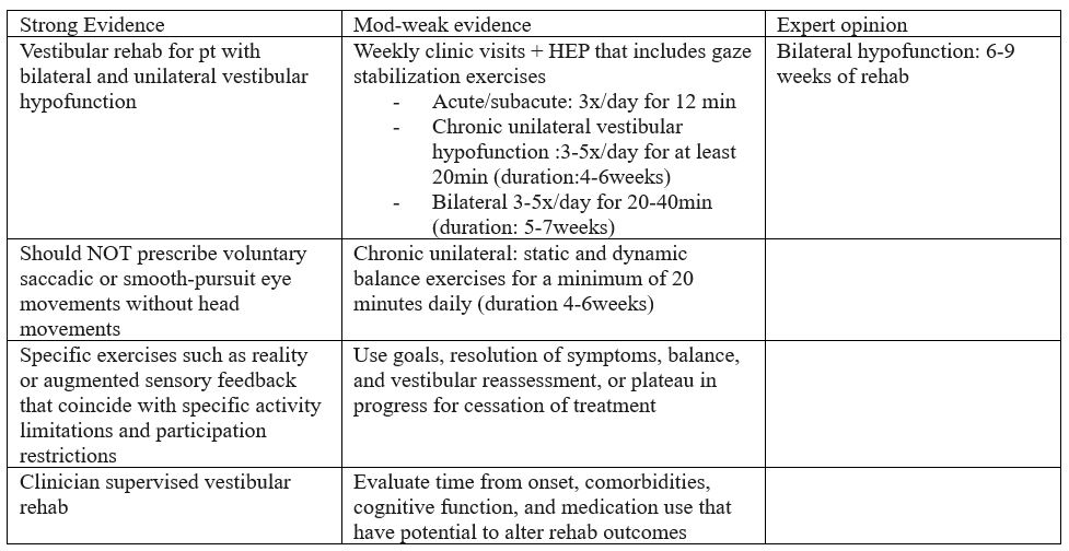
Limitations:
peripheral hypofunction (does not apply to individuals with central disorders)
References:
Hall CD, Herdman SJ, Whitney SL, et al. Vestibular Rehabilitation for Peripheral Vestibular Hypofunction: An Updated Clinical Practice Guideline From the Academy of Neurologic Physical Therapy of the American Physical Therapy Association. J Neurol Phys Ther. 2022;46(2):118-177. doi:10.1097/NPT.0000000000000382
by ptfadmin | Feb 23, 2026 | Health Tips
Reviewed by Tyler Tice, PT, DPT, OCS, ATC
This peer-reviewed article aims to identify the indications, technique, and complications of radiofrequency ablation (RFA). RFA is a minimally invasive procedure that utilizes high-frequency alternating electrical current to generate heat, resulting in targeted tissue destruction. Thermal energy is applied near or on peripheral nerve origins along the spinal cord. Parameters for temperature are designed to spare sensory and motor fibers, targeting only the dorsal root ganglia, which are responsible for transmitting ascending information, such as pain, touch, and temperature.
Indications for this procedure include chronic back and neck pain. Additionally, RFA has been used for radicular pain, discogenic pain, facetogenic pain, post-herpetic neuralgia, post-amputation pain, and post-inguinal herniorrhaphy pain. Contraindications to RFA include patient refusal, increased intracranial pressure, and local infection.
Radio frequency currents are guided via a catheter to an electrode near a nociceptive pathway to intercept pain impulses. The currents heat the surrounding tissue, and the tissue, in turn, heats the electrode. When the target temperature is reached, the current cycles on and off to maintain it.
Potential adverse effects include bleeding, infection, needle placement-induced nerve damage, and burns. Of these, the most common complication is temporary post-procedure discomfort. Other complications may involve alopecia, pigmentation changes (particularly with facial involvement), and neuropathic pain.
The duration of analgesia is dependent on the reinnervation of the targeted tissues by the proximal axons, which typically ranges from weeks to months. In conclusion, RFA should not be used as a stand-alone treatment and is not a substitute for other methods of pain management, but an adjunct treatment.
Reference:
Ray JK, Dixon B, Przkora R. Radiofrequency Ablation. [Updated 2023 Jun 12]. In: StatPearls [Internet]. Treasure Island (FL): StatPearls Publishing; 2025 Jan-. Available from: https://www.ncbi.nlm.nih.gov/books/NBK482387/
by ptfadmin | Feb 19, 2026 | Health Tips
Reviewed by Tyler Tice, PT, DPT, OCS, ATC
This systematic review evaluated the effects of core training on balance in healthy older adults 60 years of age and older. Additionally, the effect of core training on fall prevention was explored. 11 trials were included (RCTs and single-group studies) with 443 older adults from multiple countries. Intervention consisted of core training for a least four weeks. Core training was defined as “traditional core exercise and Pilates-based core programs”.
The focus of core training includes enhancing the stability, coordination, and functional capacity of the abdominals, erector spinae, and iliopsoas. According to this study the effects of core training on older adults has a greater impact on balance and gait performance compared to younger adults likely due to lower balance baseline. Specific exercises and training parameters were not explicitly detailed in the review, making the operational definitions of the interventions unclear.
Balance outcomes were categorized into the following: static, dynamic, or combination. Dynamic balance was defined as the ability to maintain stability while moving through space, regardless of foot movement. Measures used in this study to objectively gauge dynamic balance include Gait Test, Functional Reach Test, and the Timed Get Up and Go. While static balance was defined as the ability to maintain an upright posture with bilateral foot contact on the ground. This was objectively measures using the balance board test and One-Leg Stance Test.
Core training was shown to significantly improve both dynamic and static balance. Sessions greater than 45min yielded greater improvements, most notably on performance of the Timed Get Up and Go. Regarding gait outcomes, traditional core training had more efficacy than Pilates-based training.
Limitations include small number of studies and smaller sample sizes, inconsistent risk for bias across trials, minimal evidence on long-term fall reduction, and heterogeneity in intervention pools.
Overall, the article concluded that core training significantly improves balance, markedly dynamic balance in older adults and it should be integrated into fall-prevention programs. Optimal dosage and long-term effects require further study.
Reference:
Zhong Y, Guo W, Chen P, Wang Y. Effects of core training on balance performance in older adults: a systematic review and meta-analysis. Front Public Health. 2025;13:1661460. doi:10.3389/fpubh.2025.1661460
by ptfadmin | Feb 17, 2026 | Health Tips
Reviewed by John Baur, PT, DPT, OCS, CSCS, FAAOMPT
This talk frames decision‑making as a core coaching skill, not simply a by‑product of collecting more data. French situates modern strength and conditioning (S&C) inside a noisy, high‑velocity information environment where data volume grows faster than human understanding, pressing coaches to develop repeatable processes that translate numbers into training actions. The session’s aim is to help coaches identify, review, and improve strength, power, and conditioning interventions through a structured decision framework. ([NSCA TV][1])
1) The decision environment—complicated vs. complex.
French distinguishes routine, linear problems from complex scenarios characterized by many interacting elements, emergent behavior, and no single “right” answer. In complexity, the coach’s job is to simplify without being simplistic: define the problem space, isolate leading indicators, and stage decisions (e.g., “if this, then that”) so the staff can act decisively under uncertainty. The CEU quiz underscores this by defining *complexity* as “integrated order with too many elements to understand simply,” reinforcing the need for robust but practical heuristics. ([NSCA][2])
2) Time available dictates the mode of thinking.
Decision time is shaped less by the athlete’s mood than by task complexity and the cognitive mode required. When time is short and the setting is familiar, “fast” decisions anchored in practiced rules and thresholds work well; when tasks are novel or stakes are high, the coach deliberately slows the process to an analytical mode. The quiz explicitly flags task complexity and the analytical cognitive mode as the key determinants of how much time a coach needs to decide. ([NSCA][2])
3) The data deluge and bounded rationality.
French cautions that data growth is exponential while understanding is relatively linear, so chasing perfect certainty leads to analysis paralysis. Coaches work under bounded rationality—decisions are limited by the information available and the brain’s capacity to process it. The implication is to pre‑define what “good enough” evidence looks like, protect staff attention, and favor consistent, transparent rules over ad‑hoc judgment. ([NSCA][2])
4) Emotion and human behavior.
French notes that adherence, withdrawal, and performance variability are not purely rational phenomena—emotion sits at the center of human withdrawal behaviors. High‑quality coaching decisions therefore blend objective metrics with interpersonal context and communication strategies that reduce threat and increase athlete buy‑in. ([NSCA][2])
5) From diagnosis to decision: performance determinants > interventions.
When an athlete underperforms (e.g., lower‑body striking is off), the recommended pathway is not to terminate a block or ignore the signal; it’s to identify the performance determinants (strength, rate of force development, tissue tolerance, technical timing, etc.) and adjust the adaptive strategy—volume loading, constraint‑led drills, or recovery emphasis—while monitoring the response. This “determinants‑first” logic is echoed in the quiz. ([NSCA][2])
6) Build a gated profiling pipeline—strength before power.
French describes a gated approach to athlete profiling: first confirm that an athlete meets strength standards relative to their weight class; only then do they “unlock” power profiling. This keeps testing economical, protects time, and prevents advanced diagnostics from obscuring foundational deficits. The talk’s conference listing and quiz both reference this staged approach (strength ➜ power). ([NSCA][3])
7) Force–velocity balance: classify then correct.
Within the power domain, French advocates classifying athletes along the force–velocity spectrum and then programming to pull them toward balance. For a velocity‑dominant profile—what he labels an “antelope”—the corrective emphasis is to train “heavy” to raise force capacity; for a force‑dominant “gorilla,” program on the light/fast side to build velocity. This aligns with NSCA guidance on force–velocity–power profiling as a holistic lens for tailoring training. ([NSCA][2])
8) Programming architecture: general prep starts simple.
In off‑camp general preparation, dynamic strength prescriptions start with a linear loading strategy to establish rhythm, raise chronic workloads safely, and set the stage for later variation (e.g., undulating/wave loading) as camp approaches. This respects the decision principle “simple ➜ complex” and minimizes confounds while the staff is still learning how the athlete responds. The quiz anchors this point with “start with linear loading.” ([NSCA][2])
9) Contact readiness and neck strength ratios.
The session includes neck strength profiling with target flexion:extension ratios to mitigate head/neck risk in contact and collision sports. A commonly referenced target is ~1:1.5–2 (flexion:extension)—recognizing that extensors should be stronger—alongside balanced lateral flexion. Evidence over the past few years supports comprehensive neck training to improve head stabilization and potentially reduce head kinematics during impacts. ([NSCA][2])
10) Turn decisions into a repeatable operating system.
French’s practical message is to codify decision rules—thresholds for advancing from strength to power testing, clear profiles (“antelope” vs. “gorilla”), pre‑planned loading progressions, and communication sequences that account for emotion. This reduces variance between coaches, speeds up choices in complex settings, and keeps the staff focused on interventions that actually change performance, not just dashboards. ([NSCA TV][1])
Overall, the talk urges coaches to think like applied scientists: define the question, choose the smallest valid measure, decide promptly based on bounded evidence, and then observe how the athlete adapts. Iterate quickly, document decisions, and let the athlete’s response—not the prettiness of the graph—drive the next choice. ([NSCA TV][1])
- What does complexity refer to in decision making?
- A simple situation easily understood
- A condition with integrated order and too many elements to understand simply
- A lack of decision‑making structure
Answer: B
- Which factor(s) influences how much decision-making time is required?
- The athlete’s physical condition
- Task complexity and analytical cognitive mode
- The availability of data alone
Answer: B
- What is the main challenge in data growth for modern decision making?
- Data growth is linear and our ability to understand grows exponentially
- Data growth is exponential and understanding is linear
- Data growth matches the capacity to process it
Answer: B
- _________________ is central to human withdrawal behaviors?
- Emotion
- Decision making
- Logic
Answer: A
- What is the impact of “bounded rationality” in decision making?
- Enables unlimited data processing
- Focuses only on rational aspects, ignoring emotions
- Limits decision making to the available information and our ability to process that information
Answer: C
- What decision is recommended for an athlete underperforming on lower body striking?
- Terminate training
- Identify performance determinants and adjust adaptive strategies
- Ignore performance variations until you have more data
Answer: B
- During strength profiling, the athlete is being tested on whether or not they hit performance standards against _________________. If yes, then they unlock the ability to go on to power profiling.
- Weight class norms
- Upper body strength
- Lower body strength
Answer: A
- Dynamic strength prescription during off‑camp general preparation should start with a programming strategy that utilizes a __________________ approach?
- Wave loading
- Linear loading
- Reverse linear loading
Answer: B
- What should the neck flexion to extension strength ratio be for an athlete?
- 1:1
- 1:1.5–2
- 1:3
Answer: B
- What is the recommended approach to balance force and velocity if the athlete is classified as an “antelope?”
- Train on the heavy side to build strength
- Train on the light side to build velocity
- Continue programming as planned; this is a balanced athlete
Answer: A
References
French D. Effective Decision‑Making in Strength and Conditioning
. National Strength and Conditioning Association; 2023. Available at: `https://www.nsca.tv/national-conference/season:7/videos/effective-decision-making-in-strength-and-conditioning` ([NSCA TV][1])
Context on force‑velocity profiling: NSCA. Force‑Velocity‑Power Profile Characteristics. Available at: `https://www.nsca.com/education/articles/kinetic-select/force-velocity-power-profile-characteristics/` ([NSCA][4])
Neck ratio background: Sportsmith. Neck training to improve performance and injury outcomes. Published April 25, 2024. Available at: `https://www.sportsmith.co/articles/neck-training-to-improve-performance-and-injury-outcomes/` ([Sportsmith][5])
[1]: https://www.nsca.tv/national-conference/season%3A7/videos/effective-decision-making-in-strength-and-conditioning “Effective Decision-Making in Strength and Conditioning – 2023 NatCon – NSCA TV”
[2]: https://www.nsca.com/certification/ceu-quizzes/effective-decision-making-in-strength-and-conditioning/ “effective-decision-making-in-strength-and-conditioning | NSCA”
[3]: https://www.nsca.com/globalassets/events/pdf/2023/natcon/nat23-schedule-as-of-7.7.pdf?srsltid=AfmBOoq5NTDbNx-6Rj5cfre4ejQz8dcy6Ld2d3OEimg9Cv8NFlpjpDjN&utm_source=chatgpt.com “2023 National Conference | Las Vegas, NV & Online”
[4]: https://www.nsca.com/education/articles/kinetic-select/force-velocity-power-profile-characteristics/?srsltid=AfmBOop8vVCiPwx3zWKyX1rtWSKFlMFl8KfP4FbkYkNvcjp0qyzvWVub&utm_source=chatgpt.com “Force-Velocity-Power Profile Characteristics”
[5]: https://www.sportsmith.co/articles/neck-training-to-improve-performance-and-injury-outcomes/?utm_source=chatgpt.com “Neck training to improve performance and injury outcomes”
by ptfadmin | Feb 9, 2026 | Health Tips
Reviewed by John Baur, PT, DPT, OCS, CSCS, FAAOMPT
What the session is about.
Ashley Hodge’s session aims to replace “random” glute work with a structured, evidence‑informed approach that delivers hypertrophy, strength, and carryover to sport and life. She frames the talk around two big themes: 1) classifying glute exercises by how they load the hips (so you cover all functions of the glutes); and 2) balancing training variables (volume, frequency, intensity, effort, rest) to drive long‑term progress while managing fatigue. The session also calls out the most common programming mistakes and how to avoid them. ([NSCA TV][1])
Why glutes need a system.
The glute complex (gluteus maximus, medius, minimus) works in three planes: hip extension (sagittal), abduction (frontal), and external rotation (transverse). Because daily and athletic tasks demand all three, an effective program must deliberately train the different actions and strength curves—not just repeat one “favorite” exercise. Hodge formalizes this with a simple taxonomy and weekly structure so lifters get stronger from every angle rather than accumulating junk volume.
Exercise taxonomy: three ways to load the glutes.
Hodge organizes glute exercises into three categories that correspond to how resistance acts on the hips and where tension is highest across the range of motion:
- Horizontal hip extension (e.g., barbell hip thrusts, glute bridges, frog pumps, kickbacks, some back‑extension setups). These load the glutes most at lockout—short muscle lengths. They typically create less muscle damage and systemic fatigue, so they can be trained more frequently and with higher volumes (they’re great “volume drivers”). They hit both upper and lower subdivisions of the gluteus maximus well.
- Vertical hip extension (e.g., squats, lunges/split squats, good mornings, deadlifts). These challenge the glutes most in the lengthened position and therefore tend to produce greater soreness and overall fatigue. Vertical patterns are particularly effective at training the lower subdivision of the gluteus maximus. Program them intelligently (often heavier, with longer rests, and not every set to failure) to avoid recovery bottlenecks.
- Lateral/rotary glute work (e.g., machine/band hip abduction, lateral band walks, transverse‑plane cable patterns). These emphasize the glute medius (and some glute max fibers) in the frontal and transverse planes, usually at shorter muscle lengths and smaller ROMs, producing a strong “burn” (metabolic stress) without excessive systemic fatigue. They’re high‑yield accessories that round out a program and support pelvic control.
The “Rule of Thirds.”
Hodge leverages Bret Contreras’ “Rule of Thirds” as a clean, coach‑friendly template: aim for roughly one‑third vertical, one‑third horizontal, and one‑third lateral/rotary work across your weekly glute training. The same spirit applies to intensity distribution (a third heavy/low‑rep, a third moderate, a third lighter/higher‑rep) and to effort (a third to/near failure, a third close, a third well shy), giving you a balanced stimulus while keeping fatigue in check and progress sustainable. ([BC Strength][2])
Progressive overload & mind‑muscle connection (MMC).
Hodge emphasizes that hypertrophy and strength require systematic progressive overload—raising the training stress over time via more load, more reps at a given load, better technique/ROM, modestly higher volume or frequency, or slightly shorter rests. MMC—consciously focusing tension into the target muscle—can enhance activation and help many clients “find” the glutes during a movement. Used together, overload drives adaptation while MMC improves the quality of each rep.
What actually grows muscle.
Hodge revisits the three primary mechanisms of hypertrophy and their practical implications:
- Mechanical tension is the primary driver—created when muscles contract against force (the bigger tension over time, the stronger the growth signal).
- Metabolic stress (the “pump”) is useful—think higher‑rep sets, bands, abductions—but secondary to tension.
- Muscle damage happens mostly with lengthened loading and eccentrics; it likely contributes least to growth and can hinder performance if overdone. Balance all three, but bias training decisions toward creating—and progressing—mechanical tension.
Volume, frequency, and weekly layout.
Hodge notes general hypertrophy ranges from the literature (≈10–25 sets per muscle group/week), then gives a glute‑specialization template to illustrate how to “systematize” work: a high‑volume week might include ~36 total glute sets split evenly across the three categories (≈12 sets horizontal, 12 vertical, 12 lateral/rotary). Frequency can span 2–6 sessions/week based on genetics, exercise selection, load, effort, and recovery. Many lifters will thrive with ~3 dedicated glute sessions/week, while beginners can start lower and build up.
Load, reps, and rest.
Because growth can occur across rep ranges so long as sets are sufficiently challenging, Hodge recommends using a spread of intensities and reps rather than living in one zone. Practically: keep some sets heavy, some moderate, some lighter; leave a few reps in reserve on most sets; push to true failure strategically. As for rest, she suggests roughly 2–3 min between most compound sets (longer—3–6 min—when chasing PRs), and 60–90 s for isolation/abduction work, so you can repeat quality sets without accumulating junk fatigue.
Technique, individuality, and exercise choice.
Hodge encourages small “dials” to match anatomy and intent—foot stance, pelvis orientation, ROM, tempo—plus including unilateral variations each session for symmetry and control. She uses MMC and coach’s tactile/verbal cues to ensure the target muscle is doing the work. The point is not to chase novel exercises, but to extract more from the staples by executing them well and progressing them week to week.
Frequent mistakes—and fixes.
Common pitfalls include: over‑emphasizing one vector (e.g., only hip thrusts or only squats), doing too much lengthened‑position work to failure (recovery suffers), skipping lateral/rotary patterns entirely, and piling on volume without a progressive plan. The fixes flow from the system above: distribute work across vectors (“Rule of Thirds”), bias programming toward mechanical tension while using metabolic‑stress work as support, and progress deliberately rather than randomly. ([NSCA TV][1])
- At what muscle length do the glutes produce the most active force?
Answer: Slightly stretched. (Active force typically peaks around moderate lengths on the length–tension curve.)
- Why can most people tolerate relatively high volumes of glute work?
Answer: The glutes are designed to handle high workloads (locomotion/postural role), especially when much of the plan uses shorter‑length loading and accessory patterns that don’t create excessive damage.
- Recommended weekly session count for beginners?
Answer: Two sessions per week is a practical on‑ramp before progressing toward ~3+ sessions as tolerance improves. (Hodge notes glute frequency can range 2–6×/week, with many thriving around ~3×/week.)
- Which category best targets the lower subdivision of gluteus maximus?
Answer: Vertical hip extension exercises (e.g., squats, hinges).
- Most important mechanism for hypertrophy?
Answer: Mechanical tension.
- What increases metabolic stress?
Answer: Shorter rest periods (as part of higher‑rep/“pump” work).
- Which choice illustrates progressive overload?
Answer: Lifting the same load for more repetitions.
- Which matters more for hypertrophy—progressive overload or mind‑muscle connection?
Answer: Progressive overload is more important (MMC is helpful, but overload drives adaptation).
- What is the mind‑muscle connection?
Answer: Conscious, deliberate contraction/focus on the target muscle.
- Which exercise type can you usually perform more often?
Answer: Horizontal loading exercises (e.g., hip thrusts/bridges), because they emphasize shortened‑length tension and tend to cause less soreness/fatigue.
References:
[1]: https://www.nsca.tv/videos/systematizing-glute-training “Systematizing Glute Training – 2023 PTVirt – NSCA TV”
[2]: https://www.bcstrength.com/blogs/learn-with-bret-contreras/how-to-best-train-the-glutes-rule-of-thirds?srsltid=AfmBOorpV5nBJVqjT-ERgKxlOGDZgOyX1Xt_Si_j0MVF2CKvJSzjJiNh&utm_source=chatgpt.com “How To Best Train The Glutes (Rule Of Thirds)”
[3]: https://www.nsca.com/certification/ceu-quizzes/systematizing-glute-training/ “Systematizing Glute Training | NSCA”
[4]: https://www.nsca.com/education/articles/ptq/program-design-strength-hypertrophy-glute/?srsltid=AfmBOop1cn2l3JPd474kxP2oHMzMsaDlppU0KRlfKlj8MOQGejcHzeQh&utm_source=chatgpt.com “Program Design Considerations for Optimal Strength and …”
Page 3 of 48«12345...102030...»Last »