by ptfadmin | Jul 20, 2025 | Health Tips
A Brief Systematic Review and Meta analysis (Strength & Conditioning Journal, 2025)
Reviewed by John Baur, PT, DPT, OCS CSCS, FAAOMPT
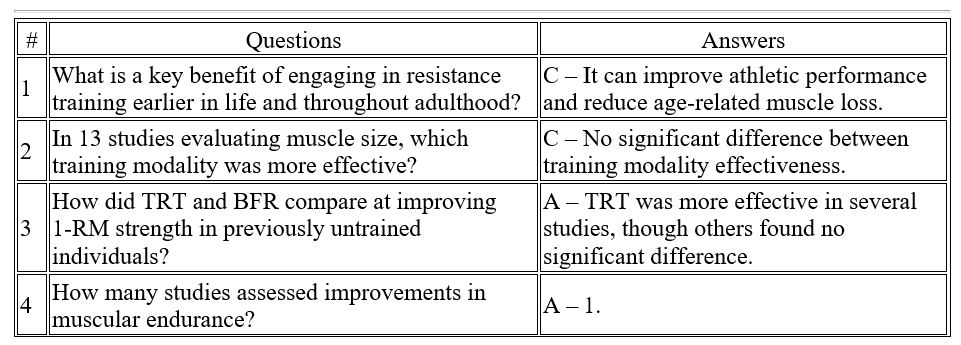
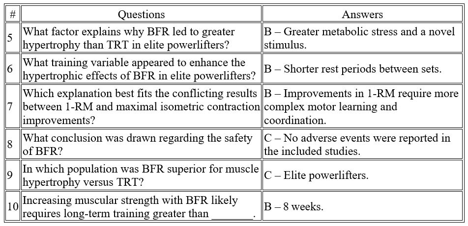
This systematic review and meta‑analysis set out to determine whether low‑load blood‑flow‑restriction (BFR) resistance training can match or outperform conventional traditional resistance training (TRT) for increasing muscle size, strength and endurance in healthy adults. Twenty randomized controlled trials (541 screened records, 20 included) met pre‑defined PICOS criteria; seven contributed hypertrophy data and nine contributed strength data to the quantitative analyses.
- Primary outcome – muscle hypertrophy: 17 of 20 trials reported significant growth with training. A pooled effect size of 0.045 (95 % CI –0.278 to 0.367) indicated no statistical difference between BFR and TRT for increases in whole‑muscle CSA or volume.
- Secondary outcome – strength: Nine studies entered the meta‑analysis (1 RM, isometric or isokinetic tests). The pooled effect size of –0.149 (95 % CI –0.439 to 0.141) likewise showed no significant difference in strength gains between modalities. Qualitative trends suggested TRT may yield faster early‑phase 1 RM gains (< 8 weeks), whereas BFR “catches up” with longer training (> 8 weeks).
- Muscle endurance: Only one study assessed endurance; both methods improved repetitions‑to‑failure equally, preventing firm conclusions.
- Contextual moderators: Rest intervals, training to failure, programme duration and athlete training status moderated results. For example, BFR out‑performed TRT for quadriceps hypertrophy in elite powerlifters—possibly due to the novel metabolic stress imposed by short‑rest, low‑load occlusion work.
- Safety profile: Across all trials no adverse events were reported, supporting BFR as a safe alternative when heavy loading is undesirable or contraindicated.
Practical takeaway: Coaches and clinicians can prescribe low‑load BFR to build muscle and strength when heavy loading is impractical, ensuring programmes run ≥ 8 weeks and use appropriately short rest intervals to maximise metabolic stress.
by ptfadmin | Jul 17, 2025 | Health Tips
Reviewed by Madison Hof, SPT
Introduction
Though conventional Total Knee Arthroplasty (TKA) procedures have yielding good long-term results in the past, robotic-assisted TKA was introduced to improve implantation alignments, particularly in younger patients. These alignment improvements were studied in this randomized controlled trial to determine if robotic-assisted TKA results demonstrate improved long-term outcome scores or implant survivorship. A large randomized, controlled trial was conducted using patients who received either robotic-assisted or conventional TKA procedures and have reached long-term (10-year minimum) follow-ups to determine if robotic-assisted TKA is superior. Terms for determination include (1) functional results based on Knee Society, WOMAC and UCLA Activity scores, (2) radiographic parameters, (3) Kaplan-Meier survivorship, and (4) complications specific to robotic-assistance, including pin tract infection, peroneal nerve palsy, pin-site fracture, or patellar complications.
Patients and Methods
1406 eligible patients were selected from one performing surgeon with surgery dates ranging from January 2002 to February 2008. 700 patients (750 knees) received robotic-assisted TKA and 706 patients (766 knees) received conventional TKA. 674 patients from the robotic-assisted TKA group and 674 patients from the conventional TKA group were available to be evaluated for clinical, radiographic, and CT scans at follow-up at an average of 13 (+/-5) years post-op. The robotic assisted TKA group consisted of 542 women and 132 men with an age range between 49 to 65 years old. The conventional TKA group consisted of 530 women and 144 men with an age range between 46 to 65 years old.
Patients from both groups were ambulatory using either crutches or a walker on the second postoperative day and were all discharged to home. Medical advice was given to all to use their assisted devices for 4 to 6 weeks and a cane thereafter as needed to prevent falls. Postoperative follow-up examinations were performed at 3 months, 1 year, and every 2 to 3 years thereafter.
Results
Functional Outcomes
It was found that there was no difference in clinical outcome measure at the most recent follow-up for those who received robotic-assisted TKAs compared to those who received conventional TKAs. Mean total Knee Society knee scores, residual pain levels, WOMAC scores, ROM, and mean UCLA activity scores showed no difference in outcomes.
Radiographic Outcomes
According to radiographic parameters measures, including limb alignment, component alignment, and aseptic loosening, there was no difference in outcomes found between groups. The rotational alignment of the femoral component from the transepicondylar axis or tibial component also showed no difference.
Component Survivorship
Both groups showed the same percentage of implant survivorship (98%) at 15 years after the operation.
Complications
The frequency of complications yielded no difference between groups. Four knees in each group acquired a superficial infection and were treated with intravenous antibiotics for 2 weeks. There were no reports of deep infection, pin site fracture, pin tract infection, patellar dislocation, patellar fracture, supracondylar fracture or peroneal nerve palsy in either group.
Discussion
It was found in this randomized controlled trial that there was no difference in outcome scores, mean implant or limb alignment, survivorship, or complications between individuals who received a robotic-assisted TKA versus individuals who received a conventional TKA at an average 10 year follow up. However, a small reduction in the proportion of knees aligned more than 3 degrees away from neutral using robotic-assisted methods was found. Meaning, there were less knees in robotic-assisted TKA that were misaligned from neutral more than 3 degrees. This demonstrates that the overall knee positioning in robotic-assisted TKA achieved more precise neutral mechanical axis alignment than conventional TKA.
Limitations of the study include having no morbidly obese patients evaluated, which is a group who may benefit more from robotic assisted procedures given the difficulties associated with identifying landmarks with a conventional TKA procedure. This study also included a population of predominantly women with low body weight and good preoperative ROM which these factors could limit the applicability of this study to other patients.
Reference
Kin YH, Yoon SH, Park JW. Does Robotic-assisted TKA Result in Better Outcome Scores or Long-Term Survivorship Than Conventional TKA? A Randomized, Controlled Trial. Clinical Orthopaedics and Related Research. 2020;478(2):266-275. Doi:https://doi.org/10.1097/corr.0000000000000916
by ptfadmin | Jul 10, 2025 | Health Tips
Reviewed by Madison Hof, SPT
Introduction
Treatment for tendinopathies should be viewed as a multimodal approach, targeting not only the peripheral tissue but the neuromuscular adaptations that occur with persistent pain as well. Addressing the corticospinal control of the muscle by motor activation as a result of excitatory and inhibitory inputs ultimately affects tendon loading and motor production. It is important to consider neuromuscular control of the asymptomatic side as well, comparing side-to-side differences as well as differences in motor control in people with and without tendinopathy.
Motor control in people with and without tendinopathy
Muscle strength is often studied in tendinopathy research as compared to motor control. It has been understood that there is no consistent pattern of strength or performance change (increase or decrease) in tendons being studied such as in Achilles, patellar and RTC tendinopathies as well as in diagnosis such as lateral epicondylalgia. Strength deficits coupled with the presence of tendinopathy has not been consistently demonstrated in past research to reasonably target the peripheral tissue’s power production alone without assessing the neuromuscular component of muscle activation.
Imbalances in recruitment ability of excitatory and inhibitory influences around the painful tendon during muscle loading were found when comparing symptomatic and asymptomatic sides of athletes with patella tendinopathy (PT). It has been hypothesized that a protective adaptation occurs, reducing the mechanical demand that is placed on the tendon. Both peripheral and central contributions are associated with motor control changes and those with PT demonstrated landing mechanic patterns with less variability than to those without PT. Motor patterns that are invariable implies that control coming from the corticospinal tract is altered and may be due to protective strategies. Jumping abilities with less variability has been shown to be a risk factor for developing PT. In fact, these individuals were shown to be better jumpers than those without PT, coining this phenomenon the ‘jumper’s knee paradox’. Therefore, movement variability has been hypothesized as an important attribute in preventing injury.
Pain is commonly accompanied with changes in motor control as an adaptive mechanism that protects us from bodily threat. Motor control while having painful symptoms is altered due to the corticospinal drive to the motor neuron or muscle. Furthermore, there were no differences in muscle strength or activation between groups suggesting that the motor task can cause a potential pain response during jumping even in the absence of nocioception.
Motor control changes may be bilateral
Side-to-side strength deficits were found to be low following unilateral Achilles tendinopathy surgery which may reflect bilateral motor control deficits. These changes following tendinopathy pain may also be system wide which can be due to intrinsic and extrinsic factors, potentially increasing the risk of tendinopathy elsewhere in the body. An increase in global sympathetic drive implies the nervous systems’ involvement in tendinopathies. For example, there is increased inhibition responses on an affected limb following a stroke and an increased corticospinal excitatory response on the unaffected limb. This indicates an increased interhemispheric inhibition from the non-affected hemisphere to the lesioned hemisphere leading to further inhibition on the affected limb during activation of the unaffected limb. Changes in the unaffected side associated with tendinopathies may be the bodies way to attempt motor control homeostasis while trying to protect a region. This may also explain the high injury prevalence of the contralateral tendon following rehabilitation.
Conclusion and clinical implications
In order to address the differences in excitability and inhibition, an alteration to corticospinal control of muscles must be acknowledged as seen in different motor strategies used for protection. Movement variability should be considered following musculoskeletal pain even if nocioceptive input is absent due to the fact that altered motor control could be transmitted to a tendon causing a continuation of previous nocioceptive input. Altered invariable movement patterns that occur with pain are presumed to optimize performance subconsciously. This is demonstrated in those with tendinopathy displaying superior strength and performance despite having inflamed peripheral tissues. Strength training is often the focus for individuals with tendinopathy to stimulate the physiological adaptation of the muscle/tendon, but it is now known that it can modulate pain and corticospinal control of the muscle as well. Rehabilitation should be considered bilaterally to address the unaffected side as well to prevent contralateral motor adaptations from developing.
References
Rio E, Kidgell D, Moseley GL, et al. Tendon neuroplastic training: changing the way we think about tendon rehabilitation: a narrative review. British Journal of Sports Medicine. 2015;50(4):209-215. doi:https://doi.org/10.1136/bjsports-2015-095215
by ptfadmin | Jul 3, 2025 | Health Tips
Introduction/Background
Carpal tunnel syndrome is a prevalent condition characterized by compression of the median nerve within the carpal tunnel pf the wrist, leading to symptoms such as numbness, tingling, and weakness in the hand. Conventional treatments include wrist splinting, corticosteroid injections, and surgical intervention. However, there is a growing interest in non-invasive, self-administered therapies that empower patients to manage their symptoms.
Methods
The double-blinded randomized controlled trial 40 participants diagnosed with mild to moderate CTS. Participants were randomly assigned to either the intervention group, which performed a specific self-stretching exercise targeting the carpal ligament, or the control group, which performed a sham stretching exercises. The intervention consisted of a daily stretching routine over a period of four weeks.
The study found that participants in the self-stretching group experienced a statistically significant improvement in symptom severity, as measured by the Boston Carpal Tunnel Questionnaire. Additionally, there was a notable increase in pinch strength among these participants compared to the control group. No adverse effects were reported, indicating the safety of the self-stretching technique.
Conclusion
The findings suggest that self-administered stretching of the carpal ligament can be an effective non-invasive treatment for reducing symptoms and enhancing pinch strength, therefore, improving hand function in individuals with CTS. This approach offers a cost-effective and accessible option for patient, potentially reducing the need for more invasive treatments. The study emphasizes the importance of patient education and adherence to the stretching regimen to achieve optimal outcomes.
References
Shem K, Wong J, Dirlikov B. Effective self-stretching of carpal ligament for the treatment of carpal tunnel syndrome: A double-blinded randomized controlled study. J Hand Ther. 2020 Jul- Sep;33(3):272-280. doi:10.1016/j.jht.2019.12.002. Epub 2020 May 1.PMID: 32362377.https://www.jhandtherapy.org/article/S0894-1130(20)30001-6/fulltext
by ptfadmin | Jun 26, 2025 | Health Tips
Reviewed by Tyler Tice, PT, DPT, OCS, ATC
Introduction/Background
The article introduces a four-stage plyometric training program as part of criterion-based rehabilitation for athletes post-ACLR. It emphasizes the importance of aligning plyometric tasks with the patient’s functional recovery status, considering factors like task intensity, momentum, ground contact time, and surface. The goal is to enhance neuromuscular function, movement quality, and reduce injury risk, facilitating a timely return to sport.
Post-ACLR, many athletes struggle to return to their previous performance levels and are at a heightened risk of re-injury. Deficits in neuromuscular performance, such as reduced strength and movement asymmetries, are common. Plyometric training involves rapid muscle lengthening followed by shortening, is highlighted as an effective method to improve explosive performance and neuromuscular control, surpassing traditional resistance training in some aspects.
This article aims to provide clinicians with a guideline on designing and implementing plyometric programs tailored to the ACLR patient’s recovery stage.
Methods
The program is divided into four stages, each aligned with specific phases of rehabilitation:
- Stage 1 (Mid-Stage Rehabilitation): Focuses on foundational movements with low intensity, emphasizing control and technique.
- Stage 2 (Late-Stage Rehabilitation): Introduces moderate-intensity exercises, incorporating more dynamic movements while maintain control.
- Stage 3 (Late-Stage Rehabilitation): Advances to higher-intensity plyometrics, emphasizing power and agility in preparation for sport-specific activities.
- Stage 4 (Return-to-Sport Training): Involves high-intensity, sport-specific plyometric exercises to simulate real-game scenarios, ensuring readiness for return to sport.
Each stage considers task intensity, movement complexity, and the athlete’s ability to perform exercises with proper technique. Progression through stages is based on meeting specific criteria, ensuring safety and effectiveness.
The article emphasizes the importance of continuous monitoring throughout the rehabilitation process. Clinicians should assess movement quality, control in both frontal and sagittal planes, and the athlete’s response to increasing exercise intensity. If an athlete cannot perform tasks with minimum competency, exercises should be simplified. Daily monitoring of pain, swelling, and soreness is recommended to guide progression and prevent setbacks.
Conclusion
Plyometric training is a crucial component of functional recovery post-ACLR. A structured, criterion-based approach ensures that exercises are matched to the athlete’s recovery status, promoting neuromuscular reconditioning and reducing the risk of re-injury. Clinicians are encouraged to integrate this four-stage program into rehabilitation protocols to optimize outcomes for ACLR patients.
References
Buckthorpe, M., and Della Villa, F. (2021). Recommendations for Plyometric Training after ACL reconstruction: A Clinical Commentary. International Journal of Sports Physical Therapy, 16(3), 879-895. https://doi.org/10.26603/001c.23549
by ptfadmin | Jun 19, 2025 | Health Tips
Reviewed by Tyler Tice, PT, DPT, OCS, ATC
Introduction/Background
Chronic low back pain is complex and often not explained by a structural pathology alone. Modern pain neuroscience suggest that persistent pain may be due to maladaptive neuroplastic changes. In this article, the authors introduce Graded Motor Imagery (GMI). GMI is a three-stage neurorehabilitation approach (left/right discrimination, explicit motor imagery, mirror therapy) designed to treat chronic pain by altering the central nervous system processing.
Methods
A 67-year-old female with a 30 year history of chronic LBP participated in the study. Her pain significantly impacted her quality of life and daily function. Prior treatments- medications, injections, physical therapy, provided limited relief. The patient was introduced to GMI as an alternative strategy.
Initial assessment revealed central sensitization, pain-related fear, and body perception disturbances. Outcomes included the Oswestry Disability Index (ODI), Pain Catastrophizing Scale (PCS), and the Fremantle Back Awareness Questionnaire (FreBAQ). The results showed high disability and distorted body perception.
GMI was introduced in three graded stages over several weeks:
- Left/Right Discrimination: The patient identified images of backs as left or right to stimulate sensorimotor processing.
- Explicit Motor Imagery: She visualized moving her back without actually moving.
- Mirror Therapy: Movements were performed with a mirror to stimulate normal movement and reduce fear and pain.
The intervention was tailored to her tolerance, emphasizing progression without exacerbating symptoms.
Results
After the GMI program, the patient reported:
- Reduced pain and disability (improved ODI scores)
- Improved body awareness (better FreBAQ scores)
- Lower fear and catastrophizing thought (reduced PCS)
She was able to return to activities she had previously avoided due to pain.
Discussion
The report highlights GMI’s potential to target central nervous system changes underlying chronic pain. It emphasizes the importance of treating not just tissue damage but also the brain’s perception of the body. The results support the integration of GMI into pain rehabilitation, particularly for patients with evidence of central sensitization and distorted body image.
Conclusion
GMI was an effective, low-cost, and non-invasive that helped reduce pain and disability in a patient with longstanding chronic LBP. The case supports its use as part of a multidisciplinary pain management approach.
References
Iglar, D., Dritsas, J., and Cortese, F. (2021). Monkey see, monkey do: Using graded motor imagery in the management of chronic low back pain- A case report. Journal of Orthopaedic & Sports Physical Therapy, 51(1), 41-41.. https://doi.org/10.2519/jospt.2021.9966